
A human chest, initially the size of a room, fills your entire view. With a few commands, you shrink it to a tiny speck, then restore it to life-size and lay it flat. You peel away the skin and layers of muscle and organs, while informative text floats in the air, projected by your headset to guide what you see.
Across the country, medical schools are increasingly using virtual reality and other digital tools to teach human anatomy. In some classrooms, screens now display detailed digital reconstructions of the human body in place of traditional cadavers, allowing students to explore bones, tendons, and muscles, watch movements in real time, and focus on specific anatomical features.
Digital Cadavers Bring Anatomy to Life in the Classroom
Sandra Brown, an occupational therapy professor at Jacksonville University in Florida, uses only digital cadavers in her introductory anatomy course. She says, “In a way, the dissection is brought to life. It’s a highly visual learning method, and students love it.”
For centuries, dissecting real human cadavers has been central to medical education, providing insight into both organ structure and the body’s interconnected systems. Being physically close to a human body has long been considered the best way to understand anatomy. Yet cadaver dissection has also faced controversy, historically tied to grave robbing and unethical practices.
Today, interactive diagrams, AI assistants, and VR experiences offer a potential alternative—no real bodies required. But as these technologies replace traditional dissection, questions remain about what might be lost and whether there are lessons from handling real human bodies that no digital tool can replicate.
The Question of Learning Beyond the Physical Cadaver
Ezra Feder, a second-year medical student at the Icahn School of Medicine at Mount Sinai in New York, reflects, “Does experiencing death provide value beyond the practical aspects of cadaver dissection? I’m not sure I have a clear answer.”
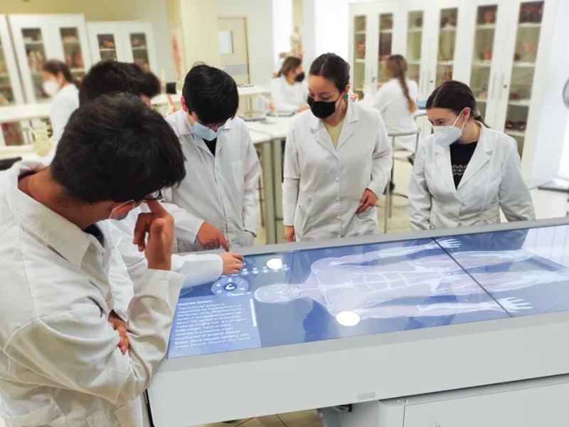
One of the most popular innovations in anatomy education is the digital cadaver “table.” Resembling oversized iPads, these portable screens can be moved easily into classrooms or labs. Anatomage, a California company behind one such table, reports that over 4,000 healthcare and educational institutions have adopted its technology. The system digitizes real cadavers into thousands of images, letting students repeatedly explore the body’s layers and systems.

Digital cadavers aren’t new, but they’ve become more realistic, interactive, and effective. Some schools have even replaced real human cadavers entirely. Brown, who teaches with the Anatomage table, notes that digital dissection aligns well with her students’ learning habits. “They’ve grown up with smartphones in their hands, so using this advanced virtual technology lets them apply the skills they already have—it was an obvious choice,” she says. “It’s really enjoyable.”
Her students can manipulate digital cadavers in ways impossible with real bodies. “They can rotate the brain upside down and view it from underneath—something you just can’t do with a fragile cadaver,” she explains. “It’s a risk-free way to explore: if they make a mistake or can’t locate something, they can simply reset or undo it.”
Other companies, such as Surglasses with its Asclepius AI Table, are advancing the digital cadaver concept even further. This table includes AI assistants with human avatars that can respond to voice commands from students and instructors, display relevant images, and quiz learners on their knowledge. Research indicates that AI assistants can enhance student learning, with avatar-based systems showing particular promise.
“Students respond well to accessible technology,” says Saeed Juggan of Yale Medical School, which provides digital anatomy tools like a 3D body model. However, Juggan cautions that AI has limitations, especially if students ask questions not covered by its data. “What happens in that case? How do you guide the bot?” he asks.
Immersive VR and AR Transform Anatomy Education
Virtual and augmented reality (VR/AR) anatomy programs have made human dissection even more futuristic. Companies like Toltech create VR headsets that let students explore detailed digital cadavers, and during the Covid-19 pandemic, Case Western Reserve students interacted with holographic bodies from home.
VR, however, has its challenges. Some students experience motion sickness with the headsets, says Kristen Ramirez of NYU’s Grossman School of Medicine. Her team’s approach is to adapt the technology to suit the specific type and content of instruction.
Ramirez and a colleague developed an in-house VR program that lets students virtually stand inside a human heart. “They can see everything a red blood cell would experience if it had eyes and cognition,” she explains.
Read the original article on: Smithsonianmag
Read more: Chinese Firm Unveils Highly Agile Life-sized Robotic Hand