
A group of UK scientists has developed a 3D embryo model that reproduces certain aspects of early human development, including blood cell formation, EFE reported Monday.
“Our model recreates the process of human fetal blood development—the blood that circulates in a baby during pregnancy—within the lab,” explained a cell biologist from the Gurdon Institute at the University of Cambridge.
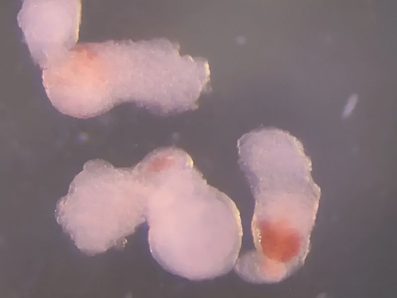
Hematoids Offer New Insights Into Early Blood Development
The hematoids, a 3D model, show “great potential” for advancing the understanding of how blood develops in the early stages of human growth.
According to University of Cambridge researchers, these new structures can also mimic diseases such as leukemia and generate long-lasting blood stem cells suitable for transplants. Stem cells are unique in their ability to divide without limit.
Published in Cell Reports, the model—designed to resemble a human embryo—recreates the cellular transformations that take place in early development, when organs and the blood system first begin to form.
From Germ Layers to Blood Formation
By the second day, the hematoids had organized into the three germ layers—ectoderm, mesoderm, and endoderm—fundamental for embryonic development. These layers “are key to forming all organs and tissues, including blood,” EFE reported. By day eight, cardiac cells appeared, marking the beginning of heart formation in a human embryo. On day 13, the researchers observed red blood spots in the hematoids, closely resembling the developmental stages of human embryos, according to the University of Cambridge.
The team also devised a method showing that hematoid-derived blood stem cells can mature into various blood cell types, including immune cells such as T cells, which defend the body against infections and abnormalities.
The university emphasized, however, that hematoids are still in the early research phase and differ significantly from real human embryos. They lack several embryonic tissues, the yolk sac that nourishes the embryo, and the placenta. Importantly, the researchers confirmed that these 3D structures cannot develop into actual embryos.
Read the original article on: Observador Pt
Read more: Rare Calico Lobster Creates a Buzz