Tag: Blood

  • This Key Blood Measurement Could Signal Your Future Disease Risk

    This Key Blood Measurement Could Signal Your Future Disease Risk

    If you've ever had a blood test ordered by a doctor, chances are it included a complete blood count (CBC). As one of the most common medical tests worldwide, CBCs are performed billions of times each year to diagnose conditions and monitor overall health.
    Credit: Pixabay

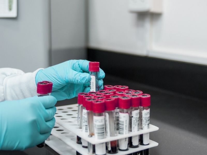
    If you’ve ever had a blood test ordered by a doctor, chances are it included a complete blood count (CBC). As one of the most common medical tests worldwide, CBCs are performed billions of times each year to diagnose conditions and monitor overall health.

    Despite their widespread use, the way clinicians interpret CBC results can often be imprecise. Currently, these tests rely on standardized reference intervals that don’t account for individual differences, which may limit their accuracy.

    At the University of Washington School of Medicine, my team and I are working to improve clinical blood testing by applying computational tools. In collaboration with the Higgins Lab at Harvard Medical School, we analyzed 20 years of blood count data from tens of thousands of patients across the U.S. Using machine learning, we developed methods to identify personalized blood count ranges and predict future disease risks.

    How CBCs and Clinical Tests Work

    Unlike purely diagnostic tests, such as those for pregnancy or COVID-19, which give a straightforward positive or negative result, most clinical tests measure biological traits that fluctuate within a regulated range.

    A CBC test provides a detailed profile of your blood, including red and white blood cell counts and platelet levels. These markers are crucial across nearly all areas of medicine. For instance:

    • Hemoglobin, an iron-containing protein in red blood cells, indicates oxygen-carrying capacity. Low levels may suggest iron deficiency.
    • Platelets, which help form blood clots, can indicate internal bleeding if their levels are too low.
    • White blood cells, essential to the immune system, often increase in response to infections.
    Lab tests are interpreted based on reference intervals.

    Redefining “Normal”

    Currently, clinicians use reference intervals based on the middle 95% of values from healthy individuals to define “normal” ranges. However, these population-based intervals don’t account for individual variability, which is largely influenced by genetics and environment.

    For example, while the standard normal platelet range is 150 to 400 billion cells per liter, your personal set point—the value your body naturally regulates—might be closer to 200, with a narrower healthy range of 150 to 250.

    This mismatch between individual set points and generalized reference intervals can lead to misdiagnoses. Doctors may overlook disease symptoms if your personal set point differs significantly from population averages or conduct unnecessary tests if your results hover near a cutoff.

    Using Machine Learning to Personalize Lab Results

    Many patients undergo routine CBCs during annual checkups, creating a rich data history. By applying machine learning to this data, we estimated blood count set points for over 50,000 patients and discovered that individual normal ranges are about three times narrower than population-based ranges. For instance, while the standard white blood cell range is 4.0 to 11.0 billion cells per liter, most individuals’ true ranges fall between 4.5 to 7.0 or 7.5 to 10.0.

    Interpreting test results based on personalized set points improved the detection of diseases like iron deficiency, chronic kidney disease, and hypothyroidism. It also allowed us to identify subtle changes that might go unnoticed when using broader reference ranges.

    Predicting Future Disease Risk

    Interestingly, individual set points also proved to be strong predictors of future health risks. For example, patients with higher white blood cell set points were more likely to develop Type 2 diabetes and faced nearly twice the risk of death from any cause compared to those with lower counts. Other blood count markers also correlated with disease and mortality risks.

    The Future of Personalized Medicine

    Incorporating personalized set points into clinical practice could revolutionize how diseases are screened and diagnosed. By leveraging your medical history to define what “healthy” truly means for you, doctors could provide more accurate and tailored care. This approach represents a promising step forward in the field of personalized medicine.


    Read Original Article: Science Alert

    Read More: Scitke

  • Blood-Derived Living Material Offers New Hope for Bone Repair, Study Finds

    Blood-Derived Living Material Offers New Hope for Bone Repair, Study Finds

    Samples of the implant material created in the lab. (University of Nottingham)

    When skin is injured, blood clots naturally initiate the healing process. Building on this mechanism, scientists have created a blood-based implant that accelerates and enhances tissue repair, particularly for broken bones.

    The research team describes this innovation as a “biocooperative regenerative” material. By incorporating synthetic peptides, the implant strengthens the blood clot’s natural barrier to improve both its structure and function. In tests on rats, the gel-like material, which can be 3D-printed, successfully repaired bone damage. If adapted for humans, this breakthrough could revolutionize healing methods.

    The ability to safely and easily transform a patient’s blood into regenerative implants is incredibly promising,” says biomedical engineer Cosimo Ligorio from the University of Nottingham. Blood is not only abundant but also easily sourced.

    The study focused on the solid regenerative hematoma (RH), a critical component in clotting. Researchers engineered peptide amphiphiles (PAs) to amplify the RH’s natural functions. These molecules enhanced clotting, linking with the RH’s scaffolding to form stronger structures.

    Enhanced Bone Repair: Modified Blood and Synthetic Peptides Activate Key Regenerative Cells

    The researchers wanted to build on the natural healing processes encouraged by blood clots. (Padilla-Lopategui et al., Advanced Materials, 2024)

    Using the modified blood combined with PAs, the team repaired small bone defects in rat skulls. Key repair cells, including mesenchymal stromal cells, endothelial cells, and fibroblasts, were activated in the implant, driving effective regeneration.

    The gel-like material could also be mechanically adjusted and 3D-printed for specific applications, showcasing its versatility.

    Regenerative medicine aims to amplify the body’s natural repair mechanisms. While effective, these processes can become overwhelmed or weakened over time, especially with age. This innovative approach could counteract those limitations, improving health and recovery outcomes.

    This ‘biocooperative’ strategy leverages natural healing mechanisms as fabrication steps to engineer materials that aid regeneration,” explains biomedical engineer Alvaro Mata from the University of Nottingham. Although still in early stages, this research highlights the potential for transformative medical applications.


    Read Original Article: Science Alert

    Read More: Scitke

  • Portable Blood Flow-Imaging Watch Observing On the Move

    Portable Blood Flow-Imaging Watch Observing On the Move

    Researchers have devised a compact version of photoacoustic imaging systems, offering a detailed view into the body, previously hindered by their bulky size. The miniaturized version fits into a watch, with its hardware housed in a backpack of comparable weight to an average five-month-old baby. This non-invasive device provides an effective means of assessing cardiac function.
    High-res photoacoustic imaging tech has been shrunk to fit in a watch
    Lei Xi/SUSTech

    Researchers have devised a compact version of photoacoustic imaging systems, offering a detailed view into the body, previously hindered by their bulky size. The miniaturized version fits into a watch, with its hardware housed in a backpack of comparable weight to an average five-month-old baby. This non-invasive device provides an effective means of assessing cardiac function.

    In simple terms, photoacoustic imaging operates as follows: Initially, an object absorbs light, such as laser pulses, converting the absorbed optical energy into heat and causing a temperature increase. Subsequently, thermoelastic expansion leads to the emission of detectable sound waves. Unlike ultrasound imaging, which primarily identifies anatomy, photoacoustic imaging provides higher-resolution functional and structural images.

    Advanced Applications of Photoacoustic Imaging

    Due to its ability to penetrate tissues up to a depth of 2-3 cm (0.8-1.2 in), photoacoustic imaging has been utilized for scanning blood vessels, estimating blood oxygenation levels (oxygen saturation), and diagnosing skin conditions and cancer. Researchers from the Southern University of Science and Technology (SUSTech) in China have developed a compact photoacoustic imaging device that can fit inside a watch.

    Lei Xi, the corresponding author of the study detailing the researchers’ new system, stated, “Although photoacoustic imaging is highly sensitive to changes in hemodynamics, challenges in miniaturizing and optimizing the imaging interface have hindered the advancement of wearable photoacoustic devices. To our knowledge, this is the first wearable photoacoustic device suitable for healthcare applications.”

    The photoacoustic watch can capture high-resolution images of blood vessels in the skin
    Lei Xi/SUSTech

    Hemodynamics refers to the dynamics of blood flow. Monitoring hemodynamic parameters such as heart rate, blood pressure, and oxygen saturation provides insight into the efficiency of cardiac function.

    The device developed by the researchers comprises a watch with an imaging interface, a handheld computer, and a backpack housing the laser and power supply (weighing 7 kg/15 lb). It is designed to allow the wearer unrestricted movement. The device’s adaptable laser focus enables imaging of multilayered structures like the skin, with a resolution of 8.7 µm adequate for imaging most small blood vessels within a maximum field of view of approximately 3 mm in diameter.

    Field Testing of Photoacoustic Device

    Volunteers utilized the photoacoustic device to evaluate its performance under various conditions, including walking and experiencing temporary blood flow blockage to the arm using a cuff. The tests confirmed that the system was functional, compact, and sufficiently stable to enable unrestricted movement.

    The backpack houses the device’s laser supply and power source and weighs 15 lb
    Lei Xi, SUSTech

    Xi highlighted the potential of miniaturized wearable imaging systems, such as the one they’ve developed, for use in community health centers to diagnose diseases initially or continuously monitor blood circulation parameters in hospitals, offering valuable insights for treating various illnesses. Further improvements could enable these systems to aid in early detection of skin conditions like psoriasis and melanoma, as well as in analyzing burns.

    The researchers are working on enhancing the system by creating a smaller laser source with a higher pulse repetition rate, which would improve compactness, reduce weight, and enhance safety and resolution, ultimately eliminating the need for the backpack.

    Xi expressed confidence in the feasibility of developing a more advanced and intelligent photoacoustic watch, leveraging rapid advancements in modern laser diode and electronic information technologies, and eliminating the reliance on a backpack.

    Additionally, the researchers aim to increase the device’s durability for strenuous physical activities like running and jumping. They also plan to incorporate more hemodynamic parameters, including qualitative assessments of blood vessel count and volume, to support early diagnosis of cancer and cardiovascular disease.


    Read the original article on: New Atlas

    Read more: Revolutionary Biorobotic Heart: A Breakthrough in Cardiac Research and Surgery

  • The Oldest Mosquito Fossils Reveal that Male Mosquitoes Fed on Blood

    The Oldest Mosquito Fossils Reveal that Male Mosquitoes Fed on Blood

    Credit: Pixaobay

    The preserved specimens of amber display distinct mouthparts that are currently found only in female individuals

    A mosquito is probably female if you swat it on your arm or neck. Only female mosquitoes feed on blood, providing the protein they need to develop eggs. Males consume plant fluids and nectar as females’ skin-piercing mouthparts are absent. It may not have always been the case, though.

    In the early Cretaceous, about 125 million years ago, at least some male mosquitoes had acute mandibles and a long appendage with tooth-like bristles, resembling the piercing features of modern females, according to research published on Monday in Current Biology.

    Earliest Fossilized Mosquitoes Confirm Blood-Feeding Behavior

    These ancient bloodsuckers were discovered by researchers interred in amber harvested in central Lebanon 15 years ago. These are the earliest fossilized mosquitoes found. The New York Times said on Monday that the discoveries provide solid proof that the earliest known mosquitoes, both male and female, sucked blood from their hosts.

    It’s possible that mosquitoes originally were bloodsuckers, the researchers speculate, rather than developing to be bloodsuckers later in evolution. If this is the case, males might have only lost capacity when blooming plants became more widespread in the Cretaceous, providing them with a food supply without worrying about being swatted.


    Read the original article on: SCIENCE

    Read more: Crocodile’s ‘Virgin Birth’ is a First for Science’s History Books