Tag: Human Cells

  • Scientists Watch Flu Viruses Enter Human Cells Live

    Scientists Watch Flu Viruses Enter Human Cells Live

    A research team from Switzerland and Japan has closely examined how the virus behaves. Using a self-developed microscopy technique, the scientists can focus on the outer surface of human cells in a Petri dish, allowing them to observe in real time the precise moment an influenza virus enters a living cell.
    Image Credits:Emma Hyde / ETH Zurich

    A research team from Switzerland and Japan has closely examined how the virus behaves. Using a self-developed microscopy technique, the scientists can focus on the outer surface of human cells in a Petri dish, allowing them to observe in real time the precise moment an influenza virus enters a living cell.

    Led by Yohei Yamauchi, Professor of Molecular Medicine at ETH Zurich, the team made a surprising discovery. Rather than remaining passive as the influenza virus nears, the cells seem to actively try to capture it. “The infection of our body’s cells is like a dance between virus and cell,” explains Yamauchi.

    How Viruses Exploit Essential Pathways

    Although cells gain no benefit from infection, the interaction appears active because the virus takes advantage of a normal cellular uptake system that the cells rely on. This system typically transports vital substances like hormones, cholesterol, and iron into the cell.

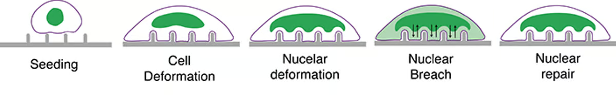
    To start the infection, an influenza virus binds to specific molecules on the cell surface. It “surfs” along the membrane, attaching to one molecule after another until it reaches an area dense with these receptors. A cluster of receptors provides the most efficient entry point.

    Once the receptors detect the virus, the membrane begins to form a small indentation at that site. A structural protein called clathrin shapes and reinforces this pocket. As the pocket deepens, it envelops the virus and forms a vesicle. The cell then pulls the vesicle inward, where the clathrin coat disassembles and releases the virus inside.

    Why Traditional Imaging Falls Short in Capturing Viral Entry

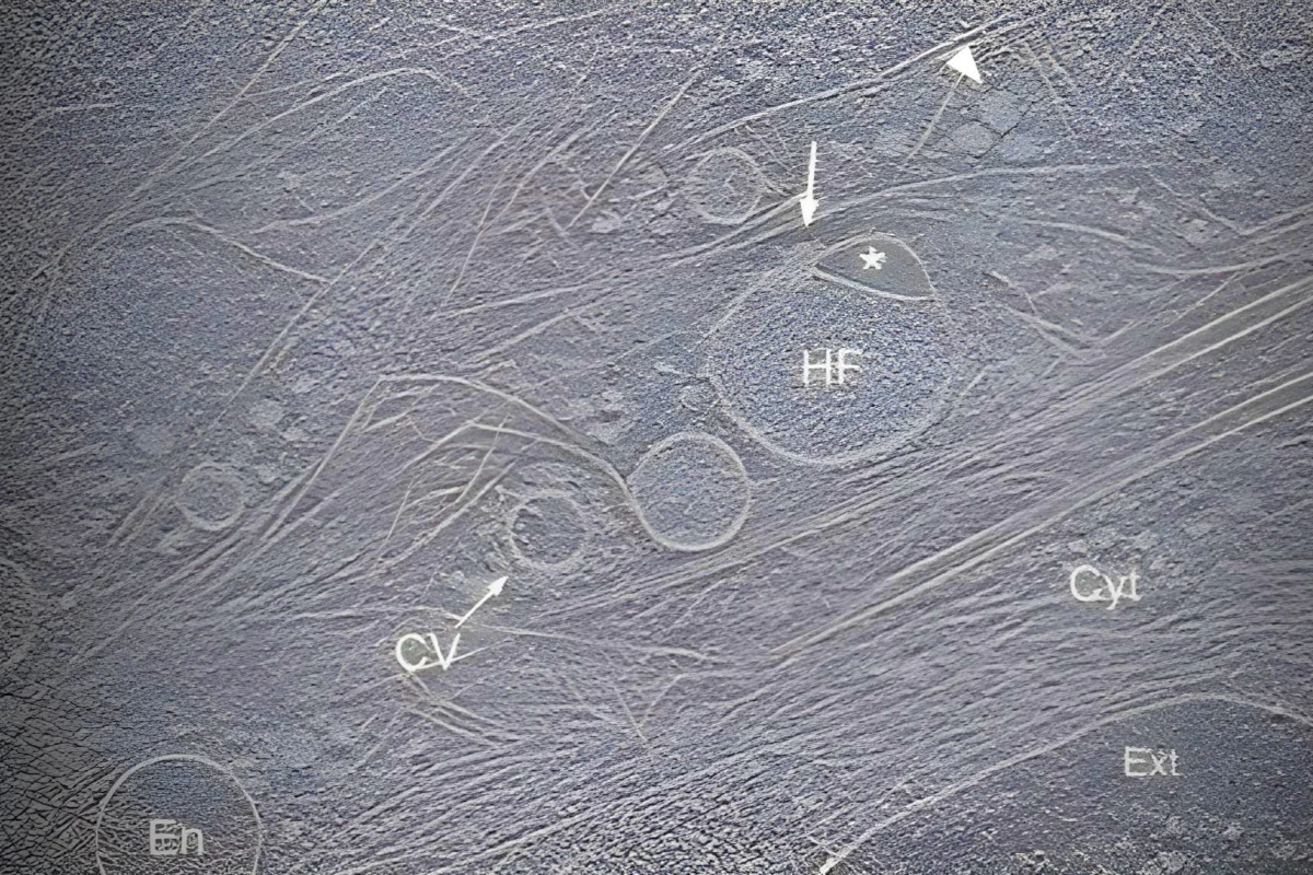
    Earlier efforts to examine this critical stage of infection depended on techniques such as electron microscopy, which destroy the cells to produce an image, capturing only isolated snapshots. Fluorescence microscopy, another frequently used method, allows live imaging but provides only low spatial resolution.

    The new technique, named virus-view dual confocal and AFM (ViViD-AFM), combines atomic force microscopy (AFM) with fluorescence microscopy. This integrated method allows researchers to observe the detailed movements of the virus as it penetrates the cell.

    Using this technique, the researchers showed that cells play an active role in multiple stages of viral entry. They recruit key clathrin proteins to the site where the virus is bound, and the membrane rises at that spot, seemingly attempting to capture the virus. These wave-like movements become stronger if the virus begins to move away from the cell surface.

    Since ViViD-AFM enables real-time observation of infection, it provides a useful method for testing potential antiviral drugs directly in cell cultures. The researchers also suggest that this technique could be applied to studying other viruses or vaccines, allowing scientists to see how these particles interact with cells as it happens.


    Read the original article on: Sciencedaily

    Read more: An Aerial Microrobot Can Reach Speeds Similar To a Bumblebee’s

  • Scientists Discover Hidden Loading Dock Structure Within Human Cells

    Scientists Discover Hidden Loading Dock Structure Within Human Cells

    Researchers at the University of Virginia (UVA) have discovered a previously unknown organelle—an ultra-small, specialized structure inside human cells that plays a role in recycling cellular material. This breakthrough could pave the way for new treatments for a variety of diseases.
    “The hemifusome is like a loading dock where they connect and transfer cargo”
    image generated using DALL-E

    Researchers at the University of Virginia (UVA) have discovered a previously unknown organelle—an ultra-small, specialized structure inside human cells that plays a role in recycling cellular material. This breakthrough could pave the way for new treatments for a variety of diseases.

    Just like our bodies rely on organs to perform vital tasks, our cells contain tiny components called organelles that carry out specific functions. Well-known examples include mitochondria, which generate energy; ribosomes, which assemble proteins; and nuclei, which house genetic information.

    UVA Scientists Uncover New Organelle, the ‘Hemifusome,’ Using Advanced 3D Imaging

    Despite decades of studying cells under microscopes, scientists at the University of Virginia have surprised the biology world with the discovery of a previously unknown organelle, which they’ve named the “hemifusome.” The team used cryo-electron tomography—a technique that rapidly freezes cells and produces detailed 3D images—to reveal this hidden structure. This method preserves biological samples in a near-natural state, allowing researchers to explore cellular interiors with exceptional clarity.

    This is like uncovering a new recycling hub within the cell,” said Seham Ebrahim of UVA’s Department of Molecular Physiology and Biological Physics. “We believe the hemifusome plays a role in organizing how cells package and handle materials. When this process breaks down, it could be linked to diseases affecting multiple body systems.”

    New Organelle Discovery May Shed Light on Alzheimer’s, Parkinson’s, and Rare Genetic Disorders

    In an interview with Virginia’s WHSV news, Ebrahim explained that the newly discovered organelle may be linked to neurodegenerative diseases like Alzheimer’s and Parkinson’s. The team also believes the finding could offer insight into rare genetic conditions such as Hermansky-Pudlak syndrome, which is associated with blood clotting issues, vision impairment, albinism, and other complications.

    We’re only starting to grasp how this organelle contributes to overall cell function and disease,” Ebrahim said. “It’s exciting because genuine cellular discoveries are rare—and this opens up an entirely new direction for research.”

    The newly discovered hemifusome, labeled “HF” in this cryo-electron tomography image
    Nature Communications (2025)

    The researchers suspect that the hemifusome plays a role in forming vesicles—small, bubble-like structures that transport substances within cells.

    Vesicles are like miniature delivery trucks,” Ebrahim explained. “The hemifusome acts as a loading dock where these trucks connect and transfer their cargo. It’s a step in the transport process we didn’t know about before.”

    Because proper sorting and movement of cellular materials is vital for cell health, studying how hemifusome disruption affects these systems could help scientists better understand the underlying causes of certain diseases and how to treat them.

    This is just the start,” Ebrahim said. “Now that we’ve identified hemifusomes, we can begin exploring how they function in healthy cells—and what goes wrong when they don’t. That knowledge might lead to new ways of tackling complex genetic disorders.”


    Read the original article on: New Atlas

    Read more: Blood Clotting Discovery Heralds A “New Era In Vascular Biology”

  • Innovative Nanopillars Pierce The Nuclei Of Human Cells

    Innovative Nanopillars Pierce The Nuclei Of Human Cells

    Researchers have discovered a way to pierce the nucleus of a cell without damaging the rest of its structure, similar to puncturing the yolk of an egg without breaking the white or the shell. This breakthrough could have significant implications for medical treatments.
    A scanning electron microscope image of a cell sitting atop the nanopillars
    Ali Sarikhani

    Researchers have discovered a way to pierce the nucleus of a cell without damaging the rest of its structure, similar to puncturing the yolk of an egg without breaking the white or the shell. This breakthrough could have significant implications for medical treatments.

    To accomplish this, scientists at the University of San Diego designed an array of nanopillars resembling blunt nails. They placed various cell types, including heart muscle, skin cells, and fibroblasts treated with fluorescent dye, on the pillars and observed the results.

    Self-Healing Nuclear Membrane

    This process created openings in the nuclear membrane while leaving the outer structure of the cells unaffected, as shown by the dye releasing from the nucleus into the cell’s cytoplasm. After the researchers removed the cells from the nanopillars, the cells closed and repaired the openings in the nuclear membrane on their own.

    This is exciting because we can selectively create tiny breaches in the nuclear membrane to access the nucleus while keeping the rest of the cell intact,” said Zeinab Jahed, the study’s senior author.

    An illustration of how the cell deforms while sitting atop the array to cause the nuclear membrane to open temporarily
    UC San Diego

    Overcoming the Nuclear Membrane Barrier

    Achieving this is no easy task, as the nuclear membrane is a notoriously tough barrier that protects our genetic code. Usually, only certain molecules can penetrate it, or a needle is used, which runs the risk of damaging the entire cell. Having a non-destructive method to access the cell nuclei could be a major advancement for gene therapy and drug delivery directly into the core of cells.

    Jahed and her team are now investigating the mechanics behind their discovery to better understand how it can be optimized for medical interventions. “Understanding these details will be key to optimizing the platform for clinical use and ensuring that it is both safe and effective for delivering genetic material into the nucleus,” she concluded.


    Read the original article: New Atlas

    Read more: All of Life’s Nucleic Acids May Have Extraterrestrial Origins