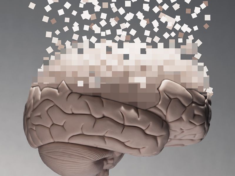

Episodic memory—the ability to remember personal experiences and past events—tends to weaken with age. While this decline is well documented, the underlying mechanisms have long been unclear. A recent study helps shed light on how and why this process occurs.
A team at the University of Oslo studied whether age-related memory loss is universal or influenced by individual risk factors like the APOE ε4 gene.
A Massive, Multi-Cohort Research Effort
Their analysis was notable for its scale. The researchers analyzed data from 3,737 healthy adults, including 10,343 MRI scans and 13,460 memory tests from multiple long-term studies.
By pooling data from dozens of cohorts, researchers have created the most detailed view yet of how age-related brain changes affect memory, says neurologist Alvaro Pascual-Leone.

The findings revealed a nuanced pattern. Although the hippocampus—a region crucial for learning and memory—played a prominent role, as anticipated, declines in memory could not be attributed to changes in any single brain region alone.
Decreases in brain tissue volume were associated with poorer episodic memory, a predictable result, but this relationship varied considerably. The link became more pronounced with advancing age, particularly after 60, and was strongest among individuals experiencing faster-than-average brain shrinkage.
The Impact of APOE ε4 on Brain Shrinkage and Memory
Participants carrying the APOE ε4 gene showed a more rapid reduction in brain tissue volume and a steeper decline in memory than others, though the overall progression followed a similar course.
According to Alvaro Pascual-Leone, cognitive and memory decline are not merely inevitable outcomes of aging, but reflect a combination of individual susceptibility and age-related biological processes that facilitate neurodegeneration and disease.
The results generate new questions while also providing important insights. Overall, they suggest memory decline is closely linked to aging, with brain changes becoming increasingly important over time.
The findings also carry implications for efforts to slow or prevent memory loss. Effective treatments will likely need to address multiple brain regions and may offer the greatest benefit if introduced early. Encouragingly, the same therapies may work for people with or without the APOE ε4 gene due to shared underlying biology.
Memory Decline Is Shaped by Multiple Interacting Factors
Evidence is mounting that memory loss later in life is shaped by a range of interacting factors within broader cognitive functioning. As researchers deepen their understanding of these influences, opportunities to manage and mitigate decline improve.
Alvaro Pascual-Leone notes that memory decline reflects broad, long-term brain vulnerability rather than a single region or gene, and understanding this could help identify at-risk individuals and develop targeted strategies to preserve cognitive health.
Read the original article on: Sciencealert
Read more:Russian Scientists Test a Plasma Engine that could Shrink Mars Travel to 30 Days