Tag: Vision

  • A light-Controlled Flexible Lens may Give Soft Machines Vision

    A light-Controlled Flexible Lens may Give Soft Machines Vision

    Inspired by the human eye, our biomedical engineering team at Georgia Tech has developed an adaptive lens made from soft, light-sensitive materials.
    Image Credits:Corey Zheng/Georgia Institute of Technology.

    Inspired by the human eye, our biomedical engineering team at Georgia Tech has developed an adaptive lens made from soft, light-sensitive materials.

    Traditional adjustable cameras rely on bulky, rigid lenses and a pupil to control focus and brightness. In contrast, the human eye achieves this through soft, flexible tissues in a compact design.

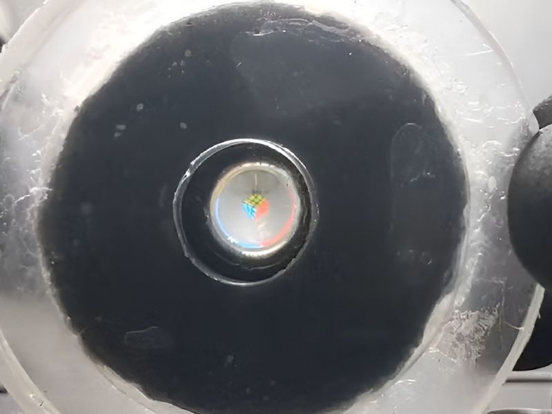
    A Soft, Light-Responsive Lens That Mimics the Human Eye

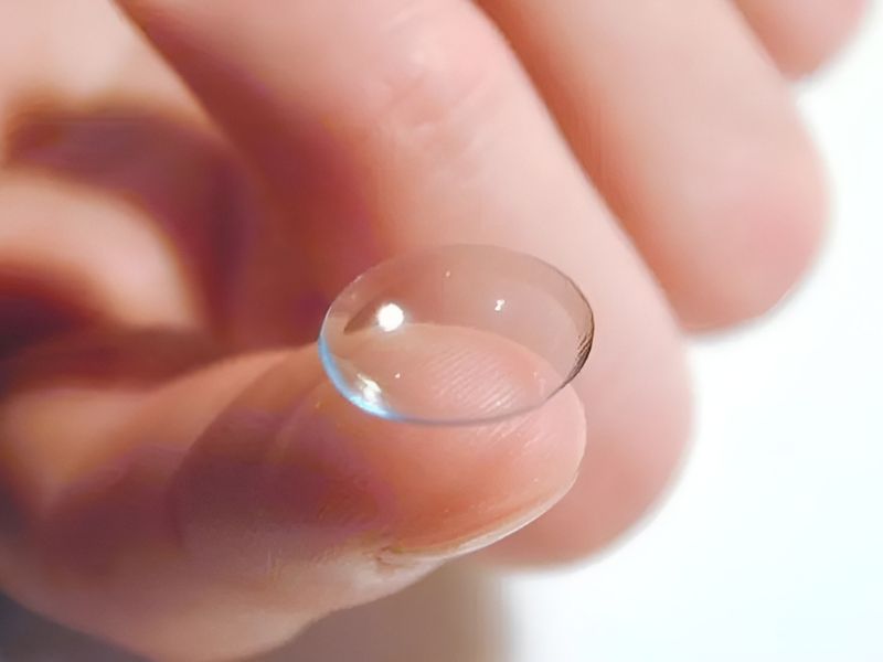
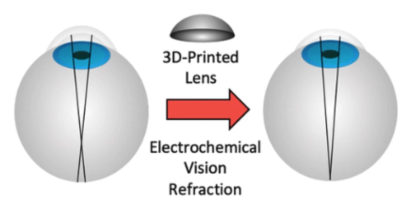
    Our innovation, the photo-responsive hydrogel soft lens (PHySL), replaces solid components with soft polymer “muscles” made from hydrogel — a water-based material. These hydrogel muscles adjust the lens’s shape to change its focal length, mimicking the action of the eye’s ciliary muscles.

    The hydrogel contracts when exposed to light, enabling contact-free control by simply projecting light onto the lens. By targeting specific areas with light, we can finely tune the lens’s shape. Without rigid parts, this flexible system is safer, more adaptable, and better suited for use with living tissue.

    Camera-based artificial vision powers many technologies, including robots and medical devices. Yet, the optical components in these systems still rely mainly on rigid, electrically powered materials. This rigidity poses challenges for emerging technologies like soft robotics and biomedical devices, which require flexible, low-power, and self-sufficient systems. Our soft lens is particularly well-suited for these applications.

    Flexible, Adaptive Machines Inspired by Nature

    Soft robots, inspired by living organisms, are built from flexible materials and structures that make them more resilient and adaptable. This approach is enabling advances in surgical endoscopes, gentle robotic grippers for handling fragile objects, and robots capable of moving through environments inaccessible to rigid machines.

    Similar principles benefit biomedical tools, where tissuelike materials create softer, safer interfaces between machines and the human body. Such materials allow devices to move naturally with the body, improving safety and comfort. Examples include skinlike wearable sensors and hydrogel-coated implants.

    image Credits:Corey Zheng/Georgia Institute of Technology.

    This research combines ideas from adjustable optics and soft “smart” materials. Although such materials are commonly used to create soft actuators—components that enable movement, like grippers or propellers—their use in optical systems has been more difficult to achieve.

    Most current soft lens designs rely on liquid-filled chambers or electronically powered actuators, which add complexity and restrict their use in fragile or wireless systems. Our light-responsive design provides a simpler, electronics-free solution.

    Advancing Performance Through Next-Generation Hydrogel Materials

    We plan to enhance our system’s performance by leveraging recent advances in hydrogel technology. Emerging studies have produced various stimuli-responsive hydrogels capable of faster and stronger contractions. By integrating these new materials, we aim to boost the functional performance of our photo-responsive hydrogel soft lens.

    We also seek to demonstrate its potential in innovative camera applications. In our current work, we created a proof-of-concept, electronics-free camera that combines our soft lens with a custom light-activated microfluidic chip. Our next step is to integrate this system into a soft robot, enabling vision without electronics. This would mark a major step forward in showcasing how our design can support new forms of soft visual sensing.


    Read the original article on: Robohub

    Read more:What is the Domain Name System? An Engineer Explains

  • Scientists Develop Contact Lenses that Offer Enhanced Vision

    Scientists Develop Contact Lenses that Offer Enhanced Vision

    Chinese researchers have created a contact lens designed to give humans a form of “super vision”—the ability to perceive infrared light, which is normally invisible to the naked eye.
    Image Credits:BBC

    Chinese researchers have created a contact lens designed to give humans a form of “super vision”—the ability to perceive infrared light, which is normally invisible to the naked eye.

    The breakthrough relies on upconversion nanoparticles, which absorb infrared light and convert it into visible light, effectively extending human vision beyond its natural 400–700 nanometer range.

    Volunteers Detect Infrared Signals Using Smart Lens

    In tests, volunteers wearing the lens could detect Morse code signals from an infrared LED and determine the light’s direction—tasks impossible without the technology.

    The lens could also prove useful in daily life, with future applications aimed at assisting people with color blindness and other vision impairments. Researchers estimate its price at around US$200 (approximately R$1,100).

    Compact, Battery-Free Lenses Surpass Traditional Night Vision Goggles

    Unlike night vision goggles—which detect infrared radiation but are bulky, require power, and display only the familiar green-tinted images—the lenses are lightweight, battery-free, and produce full-color images.

    Although still in the testing stage, the technology opens the door to a new way of seeing the world, more akin to certain animals—like birds, bees, and reindeer—that can naturally perceive invisible light bands such as ultraviolet.


    Read the original article on: G1 Globo

    Read more:The Liquid Robot Functions Similarly to a Living Cell

  • Laser-Free Vision Correction Uses Electrical Current to Reshape the Eye

    Laser-Free Vision Correction Uses Electrical Current to Reshape the Eye

    Credit: Depositphotos

    Imagine improving your eyesight in less than a minute—without lasers, scalpels, or discomfort. Scientists have unveiled a groundbreaking, non-invasive method that reshapes the cornea using only a gentle electrical current and a temporary shift in pH. Early experiments suggest it can reverse myopia without surgery, marking one of the most significant advances in vision correction since LASIK.

    Introducing Electromechanical Reshaping (EMR)

    This innovative technique, called electromechanical reshaping (EMR), modifies the cornea through low-level electrical stimulation. Researchers from Occidental College and the University of California, Irvine, described their progress during the American Chemical Society’s Fall 2025 meeting.

    The cornea—the transparent, dome-shaped layer at the front of the eye—acts as the primary lens, bending light to focus it on the retina. Composed of tightly arranged collagen fibers, it must remain smooth and strong to function properly. However, when its curve is irregular, conditions like nearsightedness (myopia) or farsightedness (hyperopia) occur.

    Illustration of EMR 
    Hill, et al./ACS Biomaterials Science & Engineering/(CC By 4.0)

    Currently, LASIK surgery is the most common option for eliminating glasses or contacts. It uses a laser to remove tiny amounts of corneal tissue beneath a flap, reshaping the surface so light focuses correctly. While LASIK boasts a high success rate—roughly 95% of patients achieve clear vision shortly after surgery—it is invasive, costly, and permanently alters the cornea’s structure.

    A New Approach to Reshaping the Eye

    Instead of cutting tissue, Hill and his colleagues explored a different approach: working with the cornea’s natural composition. Because it is largely made of collagen and charged proteins, the cornea’s shape can be temporarily softened by altering its chemical environment. By applying a mild current through a specially designed platinum “contact lens” electrode, the team shifted the tissue’s pH, making it more flexible. This brief window allowed the cornea to be reshaped inside the electrode mold.

    The electromechanical reshaping technique successfully flattened this rabbit cornea, shown in a cross section, from its original shape (white line) to a corrected one (yellow line)
    Daniel Kim and Mimi Chen

    Once the current stopped and the pH normalized, the cornea stiffened again, holding its new form. The entire procedure took about a minute, involved no cutting, and showed no signs of cell death or structural damage in lab tests. Researchers believe EMR could one day serve as an alternative to LASIK.

    An Accidental Discovery

    The discovery happened completely by accident, explained Brian Wong, professor and surgeon at UC Irvine. I was investigating how living tissues could be molded and stumbled across this chemical modification process.

    The electromechanical reshaping technique successfully flattened this rabbit cornea, shown in a cross section, from its original shape (white line) to a corrected one (yellow line)
    Daniel Kim and Mimi Chen

    To test the method, the team applied EMR to 12 rabbit corneas, successfully reshaping 10 of them to correct for myopia. Within moments, the tissue adopted the electrode’s preset curve, and early measurements confirmed proper reshaping—achieved without lasers, incisions, or trauma.

    Despite these promising results, EMR is still in the experimental phase. The technique has only been tested on isolated corneas, not live animals. Wong noted that the next step will be extensive animal trials to determine its safety, durability, and the range of corrections it can provide.

    Looking Toward Clinical Use

    There’s still a long journey from where we are now to clinical application, Hill said. But if we get there, the method could be broadly applicable, far less expensive, and potentially even reversible.

    Researchers first published details of EMR in 2023 in ACS Biomaterials Science & Engineering. Michael Hill, Daniel Kim, and Michelle Chen later presented the most recent findings at the ACS Fall 2025 meeting.


    Read the original article on: New Atlas

    Read more: Implantable Heart Patch Heals Tissue and Closes Holes

  • Breakthrough: Stem Cell Transplant Restores Vision for Multiple Patients

    Breakthrough: Stem Cell Transplant Restores Vision for Multiple Patients

    A groundbreaking stem cell transplant has greatly improved the blurred vision of three individuals with severe corneal damage.
    Credit: Pixabay

    A groundbreaking stem cell transplant has greatly improved the blurred vision of three individuals with severe corneal damage.

    This clinical trial, conducted in Japan, marks a world-first and a major milestone in stem cell research.

    Two years post-operation, no major safety issues have emerged, and all three treated corneas appear notably clearer.

    The study included four participants, each affected by limbal stem cell deficiency (LSCD), a condition that leads to scarring on the cornea.

    The Cornea’s Essential Frame

    If the cornea is viewed as the eye’s “transparent window,” then the limbus serves as its frame, holding it in place against the sclera, or white part of the eye.

    This essential frame contains a rich supply of stem cells, which act like tiny windshield wipers, replenishing damaged cells to keep the cornea clear as we age.

    Without this stem cell maintenance, gradual vision loss becomes inevitable.

    Slit-lamp microscopy images of the treated eyes. (Soma et al., The Lancet, 2024)

    When One Eye Is Affected vs. Both

    Currently, people with LSCD in one eye can have scar tissue removed and replaced with healthy corneal tissue from their other eye. However, when both eyes are affected, a donor transplant is required.

    Of the 12.7 million people worldwide with corneal vision loss, only 1 in 70 can access a transplant. Even for those who do, survival of the graft is often an issue due to the risk of rejection.

    This is where induced pluripotent stem cells (iPSCs) offer promise.

    iPSCs, created by reprogramming any human cell back to an embryonic-like state, can replicate indefinitely and transform into any adult cell type, including corneal cells.

    In 2023, U.S. researchers restored vision in two patients with corneal damage using limbic stem cells within a year of treatment.

    Now, scientists at Osaka University Hospital in Japan have advanced this work, using iPSCs derived from healthy blood cells to restore vision.

    In the lab, these iPSCs were developed into corneal epithelial cell sheets (iCEPS), which were then transplanted over patients’ corneas after scar tissue removal, with a protective contact lens placed on top.

    Seven months after the transplant, all four patients experienced improved vision. However, a year later, the vision of patient 4, a 39-year-old woman with the most severe vision impairment, had deteriorated again.

    Patients 1 and 2—a 44-year-old woman and a 66-year-old man—showed the most significant and lasting improvements.

    Potential Immune Response in Patients 3 and 4

    Researchers believe that patients 3 and 4 may not have experienced the same improvements due to an underlying immune response to the transplant. Apart from steroids, none of the patients were given immunosuppressive drugs.

    Previously, scientists used iPSCs from a patient’s own skin to restore vision for those with macular degeneration—damage to the retina’s center—but this marks the first time such an approach has succeeded in treating this particular type of vision loss without using cells from the patients themselves.

    While these small trials are promising, such procedures are still highly experimental and carry potential risks. Much more research is needed to evaluate their safety and effectiveness.

    To our knowledge, this study provides the first description of iPSC-derived cell constructs transplanted onto patients’ corneas, offering a promising future treatment option for individuals with LSCD,” the Osaka University Hospital team notes.

    To conclude, they are now preparing a multicenter clinical trial to “build on the encouraging results.”


    Read the original article on: Science Alert

    Read more: Blinking Can Enhance Your Vision, Unnoticed Until Now

  • Scientists Aid in Uncovering the Neural Mechanisms of Vision

    Scientists Aid in Uncovering the Neural Mechanisms of Vision

    When confronted with images that deviate from anticipated patterns, such as encountering a "do not enter" sign instead of the expected stop sign, how does the brain respond and adapt in contrast to exposure to images that align with predictions?
    Credit: Pixaobay

    When confronted with images that deviate from anticipated patterns, such as encountering a “do not enter” sign instead of the expected stop sign, how does the brain respond and adapt in contrast to exposure to images that align with predictions?

    A team, including researchers from York University, embarked on a quest to address a fundamental question. The prevalent theory posits that the brain constructs a predictive model of the world, and this model is adjusted when incoming sensory data contradicts it.

    However, the outcomes of the research, as detailed in the recently published paper, surprised the researchers, including Joel Zylberberg, Associate Professor at York Faculty of Science and co-corresponding author.

    Long-Term Measurement of Top-Down Signals to Sensory Areas

    According to Zylberberg, testing this theory presented challenges, necessitating the measurement of top-down signals to the brain’s sensory areas over extended periods.

    To explore how the brain learns new sensory input patterns, the researchers employed a mouse model, displaying visual patterns over multiple days and introducing images that deviated from those patterns. The focus was on the visual cortex, where retina-processed visual information is managed.

    Several researchers, including Zylberberg, are affiliated with the Canadian Institute for Advanced Research’s Learning in Machines and Brains group. This study was conducted as part of the Allen Institute for Brain Science’s Brain Observatory and OpenScope program, akin to an observatory for studying the brain through shared data, similar to astronomers collaborating to explore the universe.

    Distal Apical Dendrites Gain Sensitivity to Pattern-Violating Signals Over Time

    Measurements were taken at the distal apical dendrites and cell bodies of neurons in the visual cortex, assessing how they processed matching and pattern-violating signals. The results revealed a surprising evolution in the brain’s response to pattern-violating images over time, with distal apical dendrites becoming increasingly sensitive to such inputs, while cell bodies lost their initial strong sensitivity.

    To conclude, Zylberberg, a computational neuroscientist, notes that this discovery could provide crucial insights into sensory computation and predictive learning in the brain. The findings suggest that different forms of pattern-violating stimuli may elicit distinct prediction errors, unveiling a previously unknown component of the brain’s role in sensory learning. This understanding holds significance for improving machine learning algorithms and applications, potentially contributing to advancements in restoring vision.


    Read the original article on: Medical XPress

    Read more: Indigenous People May Have a Higher Stroke Risk

  • A Robotic Hand Employs Touch, Rather Than Vision, to Manipulate and Rotate Objects

    A Robotic Hand Employs Touch, Rather Than Vision, to Manipulate and Rotate Objects

    Scientists at the California's University San Diego have developed a novel approach, inspired by human dexterity, to enable a robotic hand to rotate objects solely through touch, eliminating the need for visual input.
    A robotic hand. Credit: Pixaobay

    Scientists at the California’s University San Diego have developed a novel approach, inspired by human dexterity, to enable a robotic hand to rotate objects solely through touch, eliminating the need for visual input.

    The team equipped a four-fingered robotic hand with 16 touch sensors on its palm and fingers. These low-cost, low-resolution touch sensors, each costing around $12, can detect whether an object is in contact with them or not, providing simple binary signals.

    The robotic hand utilizes this touch-based information to smoothly rotate a wide range of objects, including small toys, cans, fruits, and vegetables, without causing damage.

    Enabling Robots to Manipulate Objects in Low-Light and Vision-Limited Environments

    This innovative technique shows promise in enabling robots to manipulate objects in darkness or environments where visual perception is limited. The team presented their work at the 2023 Robotics: Science and Systems Conference, highlighting the potential applications of their touch-based rotational method.

    In contrast to other approaches that rely on a few high-resolution touch sensors placed at the fingertips, this method disperses many low-cost sensors across a larger area of the robotic hand, offering unique advantages and versatility.

    Xiaolong Wang, a professor specializing in electrical and computer engineering at UC San Diego and the lead researcher of this study, has pointed out several issues with current methods of robotic hand manipulation.

    Challenges in Robotic Hand Sensing and Perception

    Firstly, using a limited number of sensors on the robotic hand reduces the likelihood of contact with objects, thus restricting the system’s ability to sense its surroundings. Secondly, the complexity and cost of simulating high-resolution touch sensors that provide texture information make them impractical for real-world experiments. Lastly, many existing approaches heavily rely on visual feedback.

    To overcome these challenges, Wang and his research team propose a simple solution. They demonstrate that detailed texture information about an object is unnecessary for the task at hand. Instead, they find that binary signals indicating whether the sensors have made contact with the object or not are sufficient and much easier to simulate and implement in real-world scenarios.

    Advantages of a Comprehensive Array of Binary Touch Sensors for Robotic Object Rotation

    The researchers emphasize that using a comprehensive array of binary touch sensors provides enough data about the object’s 3D structure and orientation, enabling the robotic hand to rotate objects effectively without relying on visual cues.

    To train their system, the team utilized simulations of a virtual robotic hand manipulating various objects, including irregularly shaped ones.

    The system tracks which sensors on the hand make contact with the object during rotation, along with the positions and previous movements of the hand’s joints. Using this information, the system guides the robotic hand on the necessary joint movements for the next steps in the rotation process.

    Real-Life Testing and Object Rotation Performance

    After successful simulation training, the researchers tested the system with a physical robotic hand on unfamiliar objects. The robotic hand was able to rotate different objects, such as a tomato, pepper, a can of peanut butter, and a toy rubber duck (the most challenging due to its shape), without stalling or losing its grip. While objects with more complex shapes required more time for rotation, the robotic hand was still able to rotate them around different axes.

    In the future, Wang and his team plan to expand their approach to tackle more intricate manipulation tasks, like enabling robotic hands to catch, throw, and juggle objects. The ultimate objective is to equip robots with in-hand dexterity, a skill that comes naturally to humans but poses significant challenges for robots to master.

    Accomplishing this would greatly enhance the range of tasks that robots can perform. The research paper titled “Rotating without Seeing: Towards In-hand Dexterity through Touch” lists co-authors Binghao Huang, Yuzhe Qin, UC San Diego; and Zhao-Heng Yin and Qifeng Chen, HKUST, with the asterisk denoting equal contributions to the work.


    Read the original article on Science Daily.

    Read more: Enabling Autonomous Exploration for Robots.