Tag: Neuroscience

  • Special Gloves! Playing the Piano with Robotic Touch

    Special Gloves! Playing the Piano with Robotic Touch

    Credit: Florida Atlantic University.

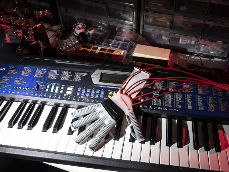
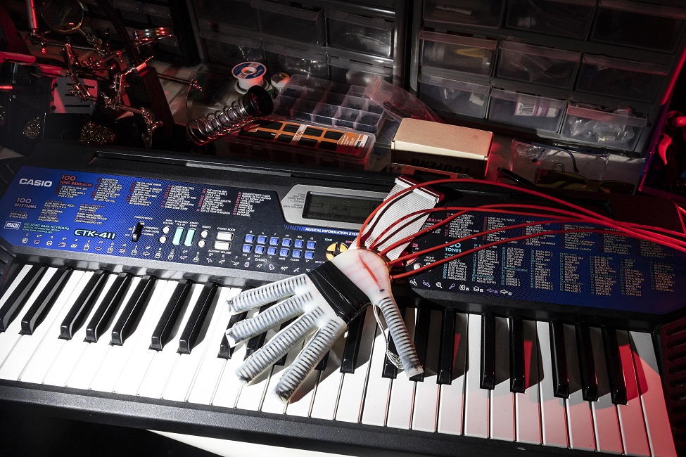
    A special glove has been created to help people who play the piano and have had a stroke. This glove is made of soft materials and uses clever technology to improve the movement of the hand. When someone has a stroke, it can be hard for them to do everyday things because their coordination and strength are affected. Robots have been made to help them, but those robots are usually stiff and not good for playing the piano.

    The new robotic glove is different because it is flexible and can “feel” if the person is playing the right notes or not. It has tiny sensors on each fingertip that can sense the movements of the hand. The glove then uses this information to give feedback and help the person play the piano better.

    The researchers tested the glove by programming it to listen to the song “Mary Had a Little Lamb” and see if it could tell when the person played the wrong notes. They made different variations of the song with mistakes, like playing the notes at the wrong time. The glove’s sensors and special algorithms helped it tell the difference between the correct and incorrect versions of the song.

    The New Gloves Results

    The results of the study showed that the glove was very good at recognizing mistakes. It could tell when someone played the wrong notes or played them at the wrong time. This is important because it means the glove can help people with disabilities relearn how to play the piano or other musical instruments.

    The robotic glove is made using a special process called 3D printing. It can be customized to fit each person’s hand perfectly. Doctors and therapists can use the information from the glove to make plans to help the person improve their weak areas. They can give them more challenging songs to practice as they get better.

    This special glove is a big breakthrough for people who have problems with their muscles and can’t use their hands properly. It’s different from other robot gloves because it can understand if the person is playing the right notes or not. Many organizations supported the research to make this glove possible, including the National Institute of Biomedical Imaging and Bioengineering and the National Science Foundation. This technology can make a big difference in helping people with disabilities regain their abilities and enjoy playing music again.


    Read the Original Article ScienceDaily.

    Read more: DeepMind Unveils Self-Training RoboCat.

  • Finding Structure in the Brain’s Static

    Finding Structure in the Brain’s Static

    Much of the electrical activity in the brain looks like noise and is not associated with reactions to any particular stimuli. The scientists found that there was structure in the noise that could reveal the state of attentiveness in the brain. In this model of brain activity in the monkey visual cortex, the overall electrical activity was measured over time in each small area. Yellow and orange areas have high activity and blue areas have low activity, corresponding to “On” and “Off” states for a set of neurons. Researchers showed a monkey images across its entire field of vision, but like most of us, the animal paid attention (attended) to only a small part of that field. When the researchers looked closely, they could see waves of activity pass over the whole visual cortex, but the waves were faster and higher in the area corresponding to the attended part of the visual field. Credit: Yan-Liang Shi/Engel lab/CSHL, 2022

    While sleeping, the whole brain flow through long, slow waves of electrical activity, like waves on a calm ocean. Scientists call that state of consciousness “slow wave sleep.” Awakening alters the pattern of electrical activity into something that resembles random noise. Yet Cold Spring Harbor Laboratory (CSHL) Assistant Professor Tatiana Engel, Postdoctoral Fellow Yianling Shi, and their collaborators found there are patterns in the noise.

    Looking at the visual processing region of a monkey brain, they found smaller, quicker, more localized versions of the large rolling sleep waves. The shapes and dynamics of these local waves connect to how attentive that part of the brain is. The researchers believe that the wave patterns offer a vital hint to comprehending anesthesia, attention, and sleep.

    The visual cortex, the part of the brain related to visual processing, is like a television screen that creates an image out of a collection of dots or “pixels.” Each brain pixel is made up of a column packed with neurons that act all together.

    Unstimulated columns flicker between being sensitive and electrically active to stimuli (” On”) or being inactive and resistant to electrical activity (” Off”). If visual information (a stimulus) strikes a visual column that is “On,” then the information is registered as a huge electrical spike. However if visual information strikes a column when it is “Off,” after that it might not be registered at all.

    Engel and Shi, in cooperation with Stanford University Professors Kwabena Boahen and Tirin Moore, and University of Washington Assistant Professor Nicholas A. Steinmetz, discovered that when monkeys are taking notice of a stimulation, the waves get much shorter and choppier. “On” and “Off” states blink through visual cortex columns driven by this stimulus faster and in a smaller area than when the animal’s attention is elsewhere.

    However, why would an attentive and awake brain intend to cycle its columns off and miss out on information? Engel has a handful of hypotheses. She claims, “keeping neurons in the ‘On’ state regularly is energetically damaging. Another reason is that if we were constantly receptive to information, we might end up being overwhelmed; the ‘Off’ state can assist subdue irrelevant information.”

    The finding that electrical noise changes patterns with different brain states might aid researchers comprehend brain responses to drugs and disease. And given that primate brains are very good at processing visual information, machine learning researchers might borrow its intelligently structured noise tricks to boost artificial brains.


    Read the original article on Medical Xpress.

    Related “DETI Brain Mapping Technique Reveals Neural Code of Vision Handling With Time”.

  • DETI Brain Mapping Technique Reveals Neural Code of Vision Handling With Time

    DETI Brain Mapping Technique Reveals Neural Code of Vision Handling With Time

    Illustration of DETI mapping of a person's brain
    DETI mapping results from the brain of a person viewing one of the stimuli used in the experiment (far left). The central column shows a flattened topographical map of the electrodes over the back of the head, illustrating the variation of DETI maps at each electrode across that scalp region. On the right-hand side, each column shows a close up of the spatiotemporal evolution of the visual code for different electrodes (each row corresponds to a different point in time in milliseconds). Each color represents one of seven different neural population responses that were mapped to each image location, thereby revealing which neural population best-coded image regions at different points in time. Credit: Bruce Hansen

    Humans are inching closer to understanding exactly how the brain codes visual information. Scientists have now established a technique that maps time-varying brain responses to images to expose just how the brain processes visual information. Colgate University Neuroscience Teacher Bruce C. Hansen worked together with Michelle R. Greene (Bates College), and also David J. Field (Cornell University) to introduce dynamic electrode-to-image (DETI) mapping. (DETI brain mapping technique).

    DETI is an analytical strategy that relies on the high temporal resolution of electroencephalography (EEG) to produce maps of visual features that are related to various neural signals with time. Observe a real-time example of neural responses mapped to an image in the video listed below.

    This video shows the neural code (at different scalp locations) for an example image. The different colors represent responses from different types of neurons. Credit: Bruce Hansen

    The study “Dynamic Electrode-to-Image (DETI) mapping shows that the human brain’s spatiotemporal code of visual details” has been published in the journal PLOS Computational Biology.

    How does the DETI brain mapping technique work?

    ” When viewing any setting, our brains code visual information across a large population of neurons in a way that enables a range of intelligent behaviors. However, the visual code used to steer behavior is not steady like a photo yet instead progresses with time and different populaces of neurons contributing to the code at different times. Our DETI mapping technique offers the first glimpse right into that time-varying code at every location in images,” said Hansen.

    Current advances in voxel-wise encoding evaluations based on functional magnetic resonance imaging (fMRI) allowed compelling reconstructions of images based upon brain data. However, they can only reproduce a single snapshot in time because of fMRI’s limited temporal resolution. The DETI mapping procedure introduced by Hansen and associates is based upon EEG signals. This allows mapping of the neural code of images with millisecond precision.

    To efficiently map the visual code to images with EEG data, Hansen and colleagues had to get over a variety of methodological obstacles. “The brain signals recorded by the EEG experience interference by the skull along with different quantities of cancellation as a result of the folding patterns of the brain.” Utilizing a biologically conceivable encoding model of the brain, Hansen and his group prevented those issues by measuring the correspondence between encoded pixels across multiple images and the resulting adjustments in the neural response.

    “One manner to think about exactly how the DETI mapping procedure functions is by running an image through the brain and projecting the subsequent neural code back onto the image.” Since EEG can measure neural signals at various scalp locations, DETI mapping creates a multiplexed view of how different populations of neurons code image attributes at different locations in images gradually. Something that was once assumed impossible to do with EEG data.

    What applications does the DETI brain mapping technique have?

    The mapping data generated by the DETI procedure uses new and vital insights into just how the neural code of images evolve in time. One of the most astounding results reported by Hansen and associates is that the brain shows up to scan images to highlight various image areas with different neural populations at other points in time. “This a scanning procedure most likely helps in an early prioritization of the ground aircraft to support judgments for navigating, with a later emphasis concentrated on sports organization.”

    These discoveries bring new and intriguing questions about how the progressing neural code notifies higher level cognitive procedures when individuals participate in different tasks. “We know that the code for visual information is scattered throughout a large population of neurons, but exactly how that code is distributed depends on the objectives of a given job. What this suggests is that the brain does not merely develop a mental picture based specifically on the environment, but instead creates a depiction that ideally matches the behavioral goals of the individual.” The excellent news is that DETI mapping makes it possible to discover the neural characteristics of task-based aesthetic codes and how those codes inevitably support task-based decision-making.


    Read the original article on Scitech Daily.

    Reference: “Dynamic Electrode-to-Image (DETI) mapping reveals the human brain’s spatiotemporal code of visual information” by Bruce C. Hansen, Michelle R. Greene and David J. Field, 27 September 2021, PLoS Computational Biology.
    DOI: 10.1371/journal.pcbi.1009456

    Related “Neurotransmitter Levels in the Brain Can Predict Math Ability”

  • Toxic Proteins Related to Parkinson’s Disease Blocked By Natural Human Molecules

    Toxic Proteins Related to Parkinson’s Disease Blocked By Natural Human Molecules

    Scientists at the UAB and the UniZar have detected a human peptide located in the brain that prevents the α-synuclein aggregates associated with Parkinson’s disease and helps prevent their neurotoxicity. The research released in Nature Communications suggests that this may be one of the organism’s natural systems to combat aggregation. The learning might help establish new therapeutic and diagnostic techniques for Parkinson’s disease and other synuclein pathologies.

    The death of neurons focused on the synthesis of dopamine, one of the brain’s primary neurotransmitters, wears away the motor and cognitive abilities of those with Parkinson’s disease. The loss of these neurons is associated with alpha-synuclein aggregation. Current studies reveal that oligomers, the initial aggregates of this protein, are the most pathogenic types of α-synuclein and account for the spreading of the disease in the brain.

    As a result, one of the more appealing strategies in combating this condition is to reduce the effects of these oligomers and, therefore, reduce pathological development. Nevertheless, the fact that these aggregates do not present a defined construct and are naturally transitory renders it incredibly challenging to detect molecules that bind with sufficient strength to explore any medical application.

    A scientific partnership between scientists from the Institute for Biotechnology and Biomedicine (IBB) at the Universitat Autònoma de Barcelona (UAB) and from the Instituto de Biocomputación y Física de Sistemas Complejos (BIFI) of the Universidad de Zaragoza (UniZar) now could detect a human endogenous peptide which strongly and particularly connects to the α-synuclein oligomers, therefore preventing their aggregation and preventing their neurotoxicity, two procedures closely related to the neurodegenerative decline of Parkinson’s disease. The recognition and research of the peptide, called LL-37, was just recently published in Nature Communications.

    Researchers explain that LL-37 engages with the toxic alpha-synuclein oligomers in a particular way and with strength above that of any peptide previously described, comparable to the strength displayed by antibodies. It hinders aggregation at a significantly reduced concentration and shields neuronal cells from being harmed.

    TLL-37 is located naturally in the human organism, both in the brain and intestine, organs in which α-synuclein aggregation occurs in Parkinson’s disease. This suggests that LL-37’s activity may react to a mechanism created by the body to naturally combat this disease, the researchers added.

    Enticed by this idea, scientists now wish to research how its expression can be controlled and if this approach can come to be a risk-free treatment with the potential to influence the course of the disease. Salvador Ventura, a researcher at the IBB and coordinator of the study, stated that there is an opportunity that a treatment for Parkinson’s disease already lies inside us and that it only requires a correct activation.

    The identification of LL-37 was carried out under the framework of research analyzing the design and features of pathogenic oligomers with the purpose of neutralizing them in a particular way. The analyses show that helical peptides with a hydrophobic side and another positively charged side are optimal for this kind of activity. The tests permitted scientists to detect three molecules with anti-aggregation activity. On top of the human molecules, a second peptide present in bacteria and a third artificially made molecule were detected.

    Along with representing a practical therapeutic option for Parkinson’s disease and other synuclein pathologies, the molecules detected in the research are encouraging resources for its diagnosis, considering that they discriminate between functional and toxic α-synuclein varieties.

    Nunilo Cremades, a researcher at BIFI-UniZar and study co-coordinator, said that until now, there were no molecules with the ability to precisely and efficiently detected toxic α-synuclein aggregates; the peptides the team presented on these issues are one-of-a-kind and, for that reason, have exceptional potential as diagnostic and prognostic resources.

    In the research, over 25,000 human peptides were computationally evaluated, and single-molecule spectroscopy techniques, along with protein engineering, were used, along with cell cultures in vitro utilizing toxic oligomers.


    Originally published by Scitechdaily.com

    Reference: “α-Helical peptidic scaffolds to target α-synuclein toxic species with nanomolar affinity” by Jaime Santos, Pablo Gracia, Susanna Navarro, Samuel Peña-Díaz, Jordi Pujols, Nunilo Cremades, Irantzu Pallarès and Salvador Ventura, 18 June 2021, Nature Communications.

  • The Risks Of Commercial Brain-Computer Interfaces

    The Risks Of Commercial Brain-Computer Interfaces

    Electroencephalography (EEG), a technique for noninvasively measuring the electrical activity of the brain
    Electroencephalography (EEG), a technique for noninvasively measuring the electrical activity of the brain. Credit: iStoock

    Researchers raise concerns about the possible social, moral, and legal repercussions of technology’s close relationship with the human brain.

    However, it may sound like a cyborg future where people can communicate with and control other people’s external technological gadgets by using their minds. Yet, this possibility may be closer than we believe.

    Brain-computer interface

    In APL Bioengineering from AIP Publishing, researchers from Imperial College London analyze contemporary commercial brain-computer interface (BCI) technologies and discuss the main technological drawbacks and ethical issues of these devices.

    Electroencephalography (EEG)

    Electroencephalography (EEG), a technique for noninvasively measuring the electrical activity of the brain, has the best chance of realizing BCI applications. Before becoming widely used, EEG-based BCIs, or eBCIs, will need to make a number of scientific advancements, but more crucially, they will cause a number of social, ethical, and legal issues.

    A schematic that demonstrates the steps a for eBCI operation. EEG sensors acquire electrical signals from the brain, which are processed and outputted to control external devices. Credit: Portillo-Lara et al.

    A few things are definite, despite the fact that it is impossible to pinpoint exactly what a user feels when using an external device with an eBCI. One is that eBCIs can converse in both directions. This not only makes it possible for someone to operate electronics, which is particularly helpful for those with disabilities who need assistance maneuvering wheelchairs, but it may also alter how the brain works.

    It has become increasingly clear that neurotechnologies have the power to significantly affect our own human interactions and sense of self, according to Rylie Green, one of the authors, who claims that for some of these patients, these gadgets have become such a fundamental component of themselves that they reject to have them removed at the conclusion of the clinical trial.

    Roberto Portillo-Lara

    In addition to these potentially negative psychological and physiological adverse reactions, intellectual property issues could allow private corporations who create eBCI technology to possess the neural data of users.

    Since neural data is frequently thought of as highly sensitive and personal data that could be linked to any given user, Roberto Portillo-Lara, another author, said that this is especially concerning. This is mostly due to the fact that, in addition to its diagnostic value, EEG data can be utilized to infer emotional and cognitive states, giving researchers unmatched insight into the intents, preferences, and feelings of users.

    Disparities in access to these technologies could exacerbate already-existing societal inequities as their availability expands beyond medical care. For instance, eBCIs can be utilized for cognitive improvement and result in severe imbalances in educational breakthroughs and professional or academic success.

    This depressing scene

    This depressing scene raises an intriguing dilemma regarding the function of legislators in the commercialization of BCI, according to Green. Should regulatory agencies step in to stop neurotechnology abuse and unequal access? Should society instead follow the example set by earlier innovations like the internet or smartphones, which were initially marketed to niche markets but are now widely used for commercial purposes?

    She urges manufacturers, producers of these technologies, future users of these technologies, and international governments to start these dialogues early and work together to find solutions to these challenging moral concerns.

    The capacity to combine the sophistication of the human intellect with the powers of contemporary technology represents an exceptional scientific achievement, according to Green, who also noted that it is starting to test our own ideas about what it means to be human.


    Originally published by: scitechdaily.com

    Reference: “Mind the gap: State-of-the-art technologies and applications for EEG-based brain-computer interfaces” by Roberto Portillo-Lara, Bogachan Tahirbegi, Christopher A.R. Chapman, Josef A. Goding and Rylie A. Green, 20 July 2021, APL Bioengineering.

    Read more: Elon Musk is Reportedly Considering Investment in Neuralink’s Rival Brain Chip Company